Related Items

TOPCON
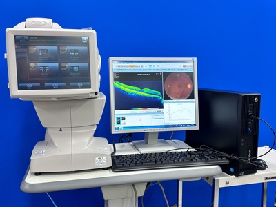
DRI OCT Triton plus
The DRI OCT Triton combines the world’s first Swept Source OCT technology with multimodal fundus imaging. Multimodal All-in-One fundus imaging tool will bring the next level of diagnostic capability to you and your patients. Combining comprehensive aspects of swept-source OCT, Topcon’s DRI OCT Triton ensures high-quality imaging. It takes into account various aspects of imaging to ensure high-resolution fundus imaging, which in turn guarantees better-quality images for more accurate assessments. These are some key features of the DRI OCT Triton that make it an ideal choice for your practice. The Topcon DRI OCT Triton is a multimodal swept-source OCT system equipped with a non-mydriatic color fundus camera. Utilizing a 1,050 nm wavelength light source, and a scanning speed of 100,000 A Scans/sec, it provides uniform scanning sensitivity allowing superior visualization of the vitreous and choroid in the same scan. Invisible OCT scanning light, eye tracking during the capture of selected scans, along with high scanning speeds reduce the effect of patient eye movement, improving workflow and allowing for more data be to collected in a shorter period. A 12 mm x 9 mm widefield scan along with automated layer segmentation provides measurement and topographical maps, with reference database, of the optic nerve and macula in one scan.

Carl Zeiss
Cirrus 4000 HD OCT
The ZEISS Cirrus HD-OCT 4000 (Cirrus HD-OCT or Cirrus) enable examination of the posterior and anterior of the eye at an extremely fine spatial scale, without surgical biopsy or even any contact with the eye. The Cirrus HD-OCT builds on and refines the retinal imaging technology first introduced with the ZEISS Stratus OCT HD-OCT stands for "high-definition optical coherence tomography."

OCT with Non-mydriatic retinal camera
TOPCON
3D OCT 1 / Maestro 1
OCT fundus tomographic images and color/red-free (digital) fundus images can be taken with one unit. Auto-alignment allows for easy and speedy imaging. Basic analysis applications are included, which are useful for diagnosing glaucoma and macular diseases. (Normative database included) Equipped with a touch panel display and a GUI design optimized for the workflow used, allowing for easy imaging. The display can be flexibly rotated, allowing imaging operations to be performed from various positions and providing better support to patients during imaging. It also provides greater freedom in installation location.

NIDEK
YC-1800
Ophthalmic YAG Laser System from NIDEK *High operability with S-switch on joystick. *Reliable laser output with unique D-pulse control. *Dual rotatable aiming beam for accurate alignment. *Wide range of focus shift. *Easily upgraded to the YAG/Green Combo system.

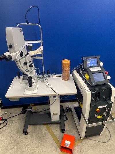
VISULAS Trion Combi YAG Laser +:532nm/561 nm/659 nm Multi color laser Combination System
Carl Zeiss
TRION II
Complete Laser Therapy System for Photodisruption and Multi-Wavelength Photocoagulation A product you can trust its capabilities will expand with your practice: perform retinal, cataract, and glaucoma therapy in one system. The VISULAS Trion Combi is the optimal system for multi-wavelength photocoagulation, post-cataract treatments, laser trabeculoplasty, and combined iridotomy. The push of a button is all you need to switch between two laser systems.

Carl Zeiss
Cirrus 500 HD-OCT Plus
The Zeiss Cirrus 500 HD-OCT "Plus" provides essential OCT capabilities with a broad range of clinical applications to comprehensive ophthalmic practices. The new OCT camera enables quick OCT fundus image refresh, making patient alignment more efficient. Main Features Cirrus 500 NEW Macular Thickness OU Analysis Ganglion Cell Analysis Guided Progression Analysis (GPA™) Macular Thickness and Change Analysis Macular Thickness Normative Data

KOWA
SL-15
The Kowa SL-15 is a highly portable, cordless, handheld slit lamp used by eye care professionals for ophthalmic examinations, featuring 10x/16x magnification, adjustable slit widths, spot illumination, a blue filter, and optional digital camera connectivity, making it ideal for domiciliary visits, emergencies, and quick room changes due to its lightweight design and rechargeable battery with a charging stand.

NIDEK
ARK-1a
The ARK-1a is a multimodality device that includes autorefraction and keratometry. This automated instrument measures with objective accuracy and features accommodation testing and retro-illumination. Super Luminescent Diode (SLD) technology allows for accurate measurements on patients with cataracts, corneal opacities, IOLs, and post LASIK procedures. The ARK-1a also utilizes a multiple pupil zone imaging method combined with rotary prism technology and an auto alignment Eye Tracking System to ensure consistent, accurate, reliable and repeatable data.

TAKARA BELMONT
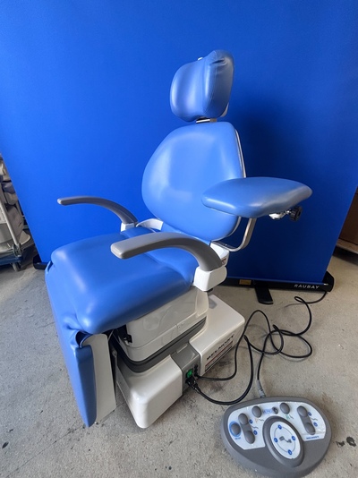
MEPRO3
This is an operating table designed specifically for ophthalmology. It is an electrically operated reclining chair. This means that it can be electrically reclined from a seated position into a supine position (surgical position), making it extremely easy to get on and off the bed. It can also be electrically raised from the surgical position back into a sitting position after surgery is completed. ■The Mepro ophthalmic operating table provides a comfortable surgical environment for all involved in surgery. ■A space-saving chair-type operating table that contributes to efficient use of the operating room. ■As it is a chair-type, it gently supports the transition from patient placement to the operating position. ■A more stable position keeps the doctor in the correct operating position.

Ophthalmic Surgical Microscope
Carl Zeiss
OPMI VISU 210 S88
Zeiss OPMI VISU 210 S88 The OPMI VISU 210 S88 ophthalmic surgical microscope for cataract and vitreo-retinal procedures boasts brilliant white light with the first of its kind Superlux Eye xenon illumination approved for ophthalmology. The OPMI VISU 210 optics incorporate the new depth of field management system allowing the surgeon to choose between brilliant red reflex or increased depth when needed. The S88 eye microscope provides superior surgeon control from the ergonomic hand grips to release the magnetic clutch brakes moving the microscope across the surgical field with minimal effort. Program the S88 with up to 9 different users with preset motorized speeds, illumination intensity and foot pedal controls.
 check!


